
Digital X-ray on site, twenty-four hours a day, with your images reviewed here rather than sent somewhere else and returned tomorrow. Most people are imaged and have an answer within the hour.
No appointment and no referral. Walk in with an injury at two in the morning and you will leave knowing whether anything is broken.
Anything dense enough to block the beam: bone, metal, glass, and air where it should not be. It is excellent at those and genuinely poor at everything soft, which is the part people are rarely told.
An X-ray is a shadow picture. Dense things cast a shadow and appear white. Soft things barely cast one at all.
What it cannot show matters just as much. Torn ligaments, damaged cartilage, muscle injuries, most soft tissue swelling, wooden splinters, and the brain are all effectively invisible on an X-ray. A normal film after a twisted ankle does not mean nothing is torn.


Yes, and this is the most useful thing on this page. Some fractures are genuinely invisible on the first X-ray and only appear seven to ten days later, once the bone has begun healing around the break.
The reason is that a hairline crack with the pieces still perfectly aligned casts almost no shadow. What makes it visible later is the healing itself, which forms new bone along the fracture line and finally gives the beam something to catch.
The wrist is where this matters most. A small bone on the thumb side of the wrist is notorious for hiding on day one, and it has a poor blood supply, so a missed break there can fail to heal properly and cause lasting problems. If you fell onto an outstretched hand and it hurts in that spot, you may be treated as though it is broken even with a normal film. That is deliberate caution, not indecision.
Hips in older adults, ribs, and the small bones of the foot behave the same way. This is why the instruction to come back if the pain has not settled in a week is a real instruction rather than a formality, and why a normal X-ray is not a reason to walk on something that still hurts two weeks later.
X-ray first for bone and chest, almost always. It is fast, cheap, low in radiation, and answers most injury questions outright. The scans exist for what it cannot see.
Think of it as the first question rather than the lesser option. For a suspected broken wrist, ankle or collarbone, an X-ray is not a compromise, it is the correct test. Sending everyone straight to a CT would add cost and radiation for no benefit.
A CT scan takes over when the question involves the head, the abdomen, blood vessels, or a fracture that an X-ray has not settled. It builds a cross-sectional picture instead of a shadow, at the cost of considerably more radiation.
Ultrasound takes over for soft tissue, pregnancy, gallbladder, and looking at blood flow, with no radiation at all. It is the better test where an X-ray would show essentially nothing.
Two X-ray views are standard, taken from different angles, because a crack invisible from the front can be obvious from the side. If you are asked to hold an awkward position for a second image, that is why.
Nothing here is a menu to choose from. Your physician picks the test that answers your question with the least radiation, and will explain the reasoning if you ask.
Very little preparation is needed, but a few things change what we do. The right-hand column is what causes repeat images, which means more radiation and a longer visit.
| Tell us or do this | Avoid this |
|---|---|
| Tell us if you are or might be pregnant | Assuming we will ask at the right moment |
| Remove jewelry, zips, and anything metal nearby | Leaving a necklace on for a chest film |
| Mention implants, plates, screws, or a pacemaker | Leaving us to guess what the metal is |
| Say if this area has been imaged before | Repeating imaging done days ago elsewhere |
| Hold still and hold your breath when asked | Moving, which blurs the image and repeats it |
| Point to exactly where it hurts most | Describing it vaguely as just the arm |
Pregnancy is the first row for a reason. An X-ray during pregnancy is often still fine, particularly for a limb, but we shield differently and sometimes choose another test entirely. Nobody will judge an uncertain answer, and it is better said early than remembered afterward.
The last row genuinely improves accuracy. Fractures hide, and the radiographer centers the image on the point you indicate. Pressing your finger on the exact spot beats a general description of the region every time.
Examined first, imaged second, and the result explained by the physician who examined you. The imaging itself takes a couple of minutes; everything around it is what takes the time.
Imaging follows an examination rather than replacing it. Where it hurts decides which images are taken.
Shielding placed where it helps, and the beam narrowed to the area being examined and no wider.
From different angles, because a fracture invisible from the front is often obvious from the side.
Digital, so there is no film to develop and no waiting. Your physician sees them almost immediately.
You see the image and have it talked through, rather than being handed a result you cannot interpret.
Splinting or immobilization if needed, referral where required, and a plan if the pain outlasts a normal film.
Very little, and the comparison most people find useful is background radiation. Everyone is exposed to a small amount every day simply from the ground, the air, and food, and a single chest X-ray is roughly what you accumulate naturally over a few days.
A limb X-ray, of a wrist or ankle, is smaller still. It is among the lowest-dose imaging in medicine, and the limb is far from the organs most sensitive to radiation.
Modern digital equipment has reduced doses substantially compared with the film systems most people remember. The beam is also narrowed to the area being examined, so the exposure is confined rather than scattered.
The honest framing is a comparison of risks. The theoretical risk from one X-ray is extremely small. The real risk of walking on an unrecognized fracture, or of missing a pneumonia, is much larger and much more immediate. Declining a needed X-ray to avoid radiation trades a tiny hypothetical harm for a substantial concrete one.
Two groups should still speak up. Tell us if you are or might be pregnant, and mention if this area has already been imaged recently elsewhere, since repeating it adds exposure for no new information. Children are imaged with settings adjusted for their size rather than adult settings.
Your images are reviewed by the physician who examined you, in the same visit. Nothing is emailed off for tomorrow.
Walk in at any hour. Imaging centers keep office hours; injuries do not.
An imaging center finds the fracture. We find it and splint it, so there is no second trip.
Rather than sending you away reassured, you are told which fractures hide and when to come back.
An X-ray here is billed as an emergency visit plus the imaging, and it is worth being clear that you cannot walk in and buy an X-ray on its own. That is not how a freestanding emergency room works, and anyone telling you otherwise is describing an imaging center.
So the honest comparison is not X-ray against X-ray. It is a full emergency assessment, imaging, interpretation and treatment in one visit, against a standalone image at an imaging center that will cost less and will not treat anything.
If you have a referral, no urgency, and simply need a picture during office hours, an imaging center is the cheaper and correct choice. We would rather say that than take a visit that did not need to happen here.
What an ER is for is the injury that just happened, out of hours, where you need to know tonight whether it is broken and to leave with it splinted. That is the value being paid for, and it is a genuinely different product.
The federal No Surprises Act protects patients from surprise bills for emergency care. Our Financial Support and Care Relief Program can substantially reduce out-of-pocket costs for those who qualify, and our Billing Disclosure page sets out the detail.
For billing questions: 817-973-7406 or info@arlingtoner.com
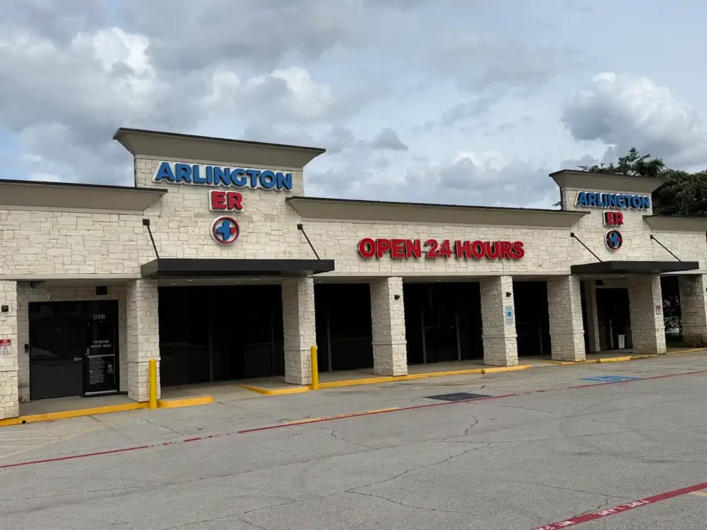
Arlington ER serves Arlington, Texas from 1607 S. Bowen Rd., on the west side of Bowen just south of Pioneer Parkway. Parking is directly outside the entrance, which matters when you are on one leg.
We are minutes from central Arlington and the Parks Mall, and a short drive from Dalworthington Gardens, Kennedale, Grand Prairie, and Mansfield. From I-20, exit at Bowen Road and head north. From Highway 303 or Pioneer Parkway, turn south onto Bowen and we are on your right. Our Contact Us page has anything else you need.
No appointment and no referral needed, at any hour of any day. If a limb is deformed, an open wound exposes bone, or the toes and fingers beyond the injury are numb or pale, call 911 rather than driving.
Digital X-ray on site twenty-four hours a day, reviewed by the physician who examined you, with treatment finished in the same visit. No appointment, no referral, no second trip. Open 24/7.
The imaging itself takes a couple of minutes. Most people are examined, imaged, given a result and treated within about an hour of arriving, since the images appear on screen within seconds of being taken.
No. Walk in and be assessed, and imaging follows if it is needed. Note that an ER cannot provide an X-ray as a standalone service — it comes as part of an emergency visit, which is a genuine difference from an imaging center.
Yes. Ask before you leave and we will arrange it. Bring them to any follow-up appointment, because a specialist comparing your original image to a later one is far more useful than describing it from memory.
The X-ray itself is completely painless and you feel nothing. What can hurt is holding an injured limb in position for a few seconds. Tell the radiographer and they will support it or find another angle.
Often yes, particularly for a limb, which is far from the abdomen and very low dose. Tell us you are or might be pregnant so we can shield appropriately or choose ultrasound instead where that answers the question.
Because a shadow picture from one angle can hide a crack completely. A fracture invisible from the front is frequently obvious from the side, so two views is the standard rather than an extra.
Come back if it has not settled within a week to ten days. Some fractures only become visible once healing has started, and a repeat image at that point often shows what the first one could not.
Metal and glass show well; wood and most plastic do not, and ultrasound is better for those. Ligaments and cartilage are invisible on X-ray entirely, so a normal film after a bad sprain does not mean nothing is torn.
Medically reviewed by Rahman Mohammed, MD | Last Updated: August 10, 2026