
Your stomach hurts, someone says the word “scan,” and you nod along without really knowing what you just agreed to. Later you wonder whether it was the right one.
It usually was, and there’s a clear reason behind the choice. Ultrasound goes first when the question is soft tissue, when you’re pregnant, or when the patient is a child. CT goes first when the question is broad and urgent.
We have both machines on site at Arlington ER in Pantego, staffed around the clock, so the decision gets made on clinical grounds rather than on whatever happens to be available at 2am.
The Short Answer: They Answer Different Questions
Ultrasound uses sound waves and no radiation. CT uses X-rays and sees almost everything in the abdomen in seconds.
Neither is better. Ultrasound wins when radiation should be avoided and the target is soft tissue or moving blood. CT wins when nobody yet knows what they’re looking for and time matters.
What Is the Real Difference Between the Two?
One uses sound. The other uses X-rays.
An ultrasound machine sends high-frequency sound into the body and builds a picture from the echoes coming back. There’s no radiation at all, it can be wheeled to the bedside, and the image updates live, so a physician can watch something move instead of studying a frozen moment.
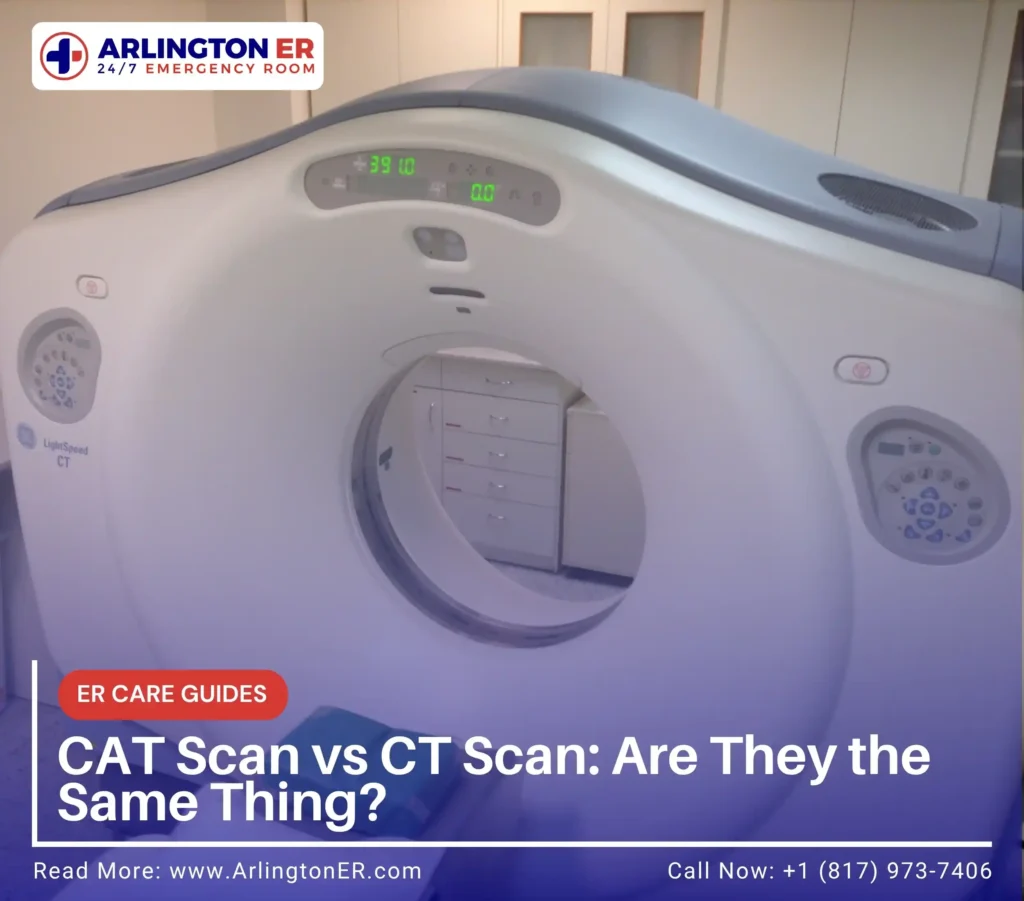
A CT takes a rapid series of X-ray images and reconstructs them into cross-sections, so the body can be read layer by layer. It’s fast, it covers a large area at once, and it doesn’t depend much on the skill of the operator or on the patient holding still. It does involve radiation. That’s the trade.
When Does Ultrasound Go First?
Whenever it can answer the question without radiation, which is more often than people assume.
- Pregnancy, at any stage, where ultrasound is usually the only imaging used
- Children, where avoiding radiation matters more because they have more years ahead in which it could matter
- Gallbladder problems, where ultrasound genuinely beats CT at showing stones
- Suspected clots in the leg, where the scan watches blood move in real time
- Testicular and ovarian pain, where the worry is blood supply being cut off
The gallbladder case surprises people who assume the more expensive scan must see more. It doesn’t. For stones in the gallbladder, sound is the better tool.
When Does CT Go First?
When the question is broad, the situation is urgent, or ultrasound has already looked and couldn’t see enough.
Severe stomach pain with no obvious source is the clearest example. Ultrasound examines one region at a time and depends on being able to see through whatever is in the way. Gas in the bowel blocks sound. A tender abdomen is hard to press on. CT has neither limitation, and images the whole abdomen and pelvis in seconds.
Major trauma is the other clear case. After a serious collision the priority is ruling out bleeding in several places at once, and there is no time to examine each region separately.
Unexplained stomach pain is one of the most common reasons people come to an emergency room, and the reasoning behind imaging it is set out in full on our page covering abdominal pain emergency care.
Which One Is Used for Each Common Problem?
| What is suspected | Usually first | Why |
| Gallbladder stones | Ultrasound | Better than CT at showing stones, and no radiation |
| Kidney stones | CT | Shows size and exact position, which decides treatment |
| Appendicitis in an adult | CT | Reliable regardless of body type or bowel gas |
| Appendicitis in a child | Ultrasound | Radiation avoidance, CT only if the view is unclear |
| Any imaging in pregnancy | Ultrasound | No radiation reaches the pregnancy |
| Suspected clot in the leg | Ultrasound | Shows blood moving, live |
| Serious trauma | CT | Covers several regions at once, in seconds |
| Unexplained severe stomach pain | CT | Broad view when the source is unknown |
Is Ultrasound Safer Than a CT Scan?
Ultrasound carries no known radiation risk, so in that narrow sense, yes. The fuller answer is that the safest scan is the one that answers the question.
A single CT of the abdomen delivers a radiation dose in the range of a few years of ordinary background exposure. That matters, and it’s why nobody orders one without a reason. It’s also small next to the risk of missing internal bleeding, a blocked kidney, or a surgical problem worsening unnoticed.
The real risk isn’t the scan. It’s the wrong scan, or one that cannot see what needs seeing, followed by a delay while everyone waits to find out.
Can You Ask for One Instead of the Other?

You can always ask, and you should. Two questions get you most of the way: what are we looking for, and would ultrasound answer this?
Sometimes the answer is yes and the plan changes. Often it’s no, for a specific reason, and hearing that reason is usually more reassuring than the scan itself.
If you’re pregnant, or think you might be, say so before any imaging. Every time, even if you have already mentioned it. That one sentence changes the whole approach.
What Happens After the Scan?
An emergency physician reads it and acts on it during the same visit. That’s the difference between an emergency room and an imaging appointment: nobody sends you home to wait for a call.
Imaging rarely stands alone either. Bloodwork usually runs alongside it, because a picture shows structure while blood shows function. All of it runs here through on-site lab testing rather than being sent elsewhere.
Where a scan is needed, what the CT involves is set out on our page covering emergency CT scan services, and the same detail for sound-based medical imaging sits on our emergency ultrasound services page.
Frequently Asked Questions
Sometimes, and it’s the preferred first test in children and in pregnancy for that reason. In adults it’s less reliable, because bowel gas and body type can block the view. When ultrasound looks and cannot give a clear answer, CT is the usual next step.
Because the ultrasound couldn’t see enough to settle the question. That’s a normal sequence, not a sign something went wrong. Starting with the test that carries no radiation, then escalating only when it cannot answer, is deliberate.
No. A warm gel is applied and a probe is moved across the skin with light pressure. If the area is already tender, that pressure can be briefly uncomfortable, but the scan itself does nothing painful.
A CT takes seconds once you are on the table. An ultrasound usually takes longer, because the person performing it is hunting for specific views. In both cases the assessment beforehand and the interpretation afterward take longer than the scan.
It can find masses and abnormalities that need further investigation, but it cannot diagnose cancer on its own. That takes additional imaging and usually tissue sampling. An emergency room’s job is to identify what needs urgent attention and make sure the right follow-up happens.